Selamat datang di dunia judi online! Bagi Anda yang mencari hiburan dan kesenangan, permainan seperti Nenektogel4D, casino, slot, sbobet, live ball, poker, togel sgp, togel hk, dan togel sdy merupakan pilihan yang tepat. Semua permainan ini menawarkan keseruan dan peluang menang yang menarik. Apakah Anda seorang pemula atau sudah berpengalaman, ada banyak opsi untuk Anda nikmati. Mari kita jelajahi lebih dalam tentang permainan-permainan yang menarik ini!
Berbicara mengenai judi online, perlu diingat bahwa togel telah menjadi salah satu permainan yang sangat populer, khususnya togel sdy, togel hk, dan togel sgp. Dengan mengandalkan keberuntungan dan rumus tertentu, pemain dapat memprediksi angka-angka yang akan keluar, dengan harapan bisa memenangkan hadiah besar. Bagi Anda yang suka tantangan dan memiliki strategi yang baik, Nenektogel4D mungkin bisa menjadi pilihan yang menarik.
Namun, jika Anda lebih suka permainan kasino klasik seperti roulette, blackjack, atau mesin slot, Anda juga dapat menikmatinya secara online. Casino online menawarkan pengalaman serupa dengan keuntungan tambahan berupa akses 24/7 dan beragam pilihan permainan. Mulai dari pemula hingga pemain berpengalaman, ada meja dan mesin slot untuk setiap tingkatan.
Selain itu, jika Anda menyukai pertaruhan olahraga, sbobet menyediakan platform taruhan olahraga online yang menarik. Dari sepak bola hingga balap kuda, Anda dapat memasang taruhan pada tim atau atlet favorit Anda. Sementara itu, untuk penggemar poker, permainan ini telah menjadi favorit selama bertahun-tahun. Online poker menyediakan ruang virtual untuk bermain dan bersaing dengan pemain lain di seluruh dunia.
Dengan begitu banyak pilihan permainan yang tersedia di dunia judi online, ada sesuatu untuk semua orang. Apakah Anda mencari kesenangan, hiburan, atau keuntungan finansial, judi online dapat memenuhi semua kebutuhan Anda. Ini adalah waktu yang menarik untuk menjelajahi dunia judi online dan menguji keberuntungan Anda. Siapkan strategi Anda, pasang taruhan Anda, dan nikmati keseruan judi online di kenyamanan rumah Anda sendiri. Selamat bermain dan semoga beruntung!
Togel Online
Togel online adalah bentuk perjudian yang populer di kalangan penjudi daring. Ada berbagai jenis togel yang dapat dimainkan, seperti togel sdy, togel hk, dan togel sgp. Dalam permainan togel online, pemain harus menebak angka yang akan keluar pada hasil undian tertentu. Togel sdy, togel hk, dan togel sgp adalah variasi togel yang terkenal dan banyak diminati.
Nenektogel4D adalah salah satu situs judi online yang menawarkan permainan togel online. Melalui situs ini, pemain dapat memasang taruhan pada togel sdy, togel hk, dan togel sgp dengan mudah dan aman. Nenektogel4D menyediakan berbagai pasaran togel, sehingga pemain memiliki banyak pilihan untuk memasang taruhan dan meningkatkan peluang kemenangan mereka.
Bermain togel online juga memberikan kemudahan akses. notinmymarinecorps dapat mengakses permainan ini kapan saja dan di mana saja melalui perangkat komputer atau ponsel cerdas. Selain itu, togel online juga menawarkan berbagai macam metode pembayaran yang aman dan nyaman. Pemain dapat melakukan setoran dan penarikan secara online, tanpa perlu ke kasino fisik atau agen togel offline.
Dalam togel online, keberuntungan dan strategi berperan penting. Pemain perlu melakukan analisis statistik dan memperhatikan pola angka yang sering muncul. Dengan memahami karakteristik togel sdy, togel hk, dan togel sgp, pemain dapat meningkatkan peluang kemenangan mereka. Namun, penting untuk diingat bahwa togel online bersifat acak dan hasilnya tidak dapat diprediksi dengan pasti.
Permainan Casino Online
Permainan di casino online menawarkan pengalaman judi yang seru dan mengasyikkan bagi para penggemar taruhan. Di sini, pemain dapat menikmati berbagai jenis permainan seperti roulette, blackjack, dan baccarat. Terlebih lagi, ada juga variasi permainan seperti sic bo, dragon tiger, dan masih banyak lagi yang menghadirkan sensasi seru di setiap putaran.
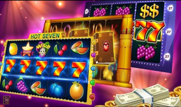
Salah satu permainan yang tidak boleh dilewatkan adalah slot online. Permainan ini menampilkan berbagai tema menarik dan memiliki banyak hadiah menanti. Dari tema klasik hingga tema populer seperti film dan musik, setiap mesin slot memiliki karakteristik yang unik. Pemain dapat memutar gulungan dan berharap mendapatkan kombinasi simbol yang menguntungkan untuk memenangkan hadiah besar.
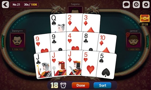
Selain itu, permainan kartu seperti poker juga sangat populer di dunia casino online. Dalam permainan poker, pemain akan bersaing dengan pemain lain untuk mencapai kombinasi kartu terbaik dan memenangkan pot. Ada berbagai variasi poker yang bisa dimainkan, termasuk Texas Hold’em, Omaha, dan Seven-Card Stud. Permainan ini bisa dimainkan dalam mode turnamen atau meja langsung dengan pemain lain di seluruh dunia.
Dengan kemajuan teknologi, casino online juga menyediakan opsi taruhan langsung seperti live ball. Pemain dapat memasang taruhan pada pertandingan olahraga yang sedang berlangsung secara real-time. Hal ini memberikan kesempatan kepada pemain untuk merasakan sensasi taruhan langsung dan mendukung tim favorit mereka. Dalam live ball, pemain dapat memasang taruhan pada berbagai jenis olahraga populer seperti sepak bola, basket, tenis, dan masih banyak lagi.
Itulah beberapa jenis permainan casino online yang bisa dinikmati oleh para penggemar judi. Dari slot yang menarik, poker yang menantang, hingga taruhan langsung di pertandingan olahraga, casino online menyediakan hiburan yang tak terbatas dan peluang untuk memenangkan hadiah besar.
Judi Online Lainnya
Berbicara tentang judi online, kita juga tidak bisa melupakan beberapa varian permainan yang populer di dunia perjudian digital seperti poker, casino, dan taruhan bola live.
Poker adalah permainan kartu yang terkenal di dunia judi online. Banyak pemain yang merasa tertantang dengan strategi pemain lain dan keberuntungan dalam permainan ini. Di situs-situs judi online, Anda dapat menemukan berbagai jenis poker seperti Texas Hold’em, Omaha, dan 7-Card Stud.
Selanjutnya, casino online juga merupakan pilihan yang menarik bagi pecinta judi online. Anda dapat menikmati beragam permainan seperti blackjack, roulette, baccarat, dan slot. Casino online memungkinkan Anda untuk merasakan atmosfer kasino sungguhan tanpa harus pergi ke tempat fisiknya.
Tidak hanya itu, taruhan bola live juga sangat diminati para penggemar olahraga dan pemain judi online. Anda dapat memasang taruhan pada pertandingan sepak bola langsung dan menonton pertandingannya secara live di situs judi online. Dengan begitu, Anda dapat merasakan sensasi mendukung tim favorit Anda sambil mendapatkan peluang untuk memenangkan taruhan Anda.
Itulah beberapa jenis judi online yang populer di dunia perjudian digital. Setiap permainan menawarkan pengalaman berbeda dan keasyikan tersendiri bagi para pemainnya. Jadi, tunggu apa lagi? Mulai jelajahi dunia judi online sekarang dan rasakan keseruan serta kemenangan yang menggiurkan!